Oh No, I Discovered A Lump On My Pet! Part II
| Tweet |
Last time, I discussed three very common lumps I come across in my canine patients. There are a myriad of lumps and bumps that can affect your pets and I simply can’t cover all of them. I do, however, want to shed some light on a few other prevalent lumps and just equip you with an appropriate strategy to tackle any lump your pet may develop.
I must warn you, there will be lots of graphic pictures included in this post!
These are generally secondary to a malformation of the hair follicle or as I like to refer to it as a blogged up hair follicle or pore. They lead to hard oval shaped lumps under the skin. They are usually not painful. However, if they grow too rapidly, they can burst and develop secondary infection and irritate your pet. A fine needle aspirate usually yields a very thick yellowish material (sebum: oily secretion produced by sebaceous gland) and examination under the microscope will allow for a definitive diagnosis.
It is best to leave them alone and monitor them.If they start bothering your pet, then they should be surgically removed.
I have sometimes incised them under local anesthetic to express their content and chemically cauterize their inner lining. However, I always put these patients on a course of antibiotics and explain to their owners that this is only a band aid solution as they will most likely recur if they are not surgically removed.
They can often look like this and mostly occur around the head, chest or back of your pet.
These lumps can arise from cartilage, nerves, fat or even vessels. They usually feel quite solid and appear to have a very distinct capsule.
Unfortunately this type of tumor doesn’t aspirate well and so fine needle aspirates will often give us misleading results.
For example, this is a fine needle aspirate from a hard lump that grew suddenly on Chloe. At the time, I didn’t know it was a sarcoma based on this slide. However, I recommended removal of the lump either way due to its rapid growth.
As you can observe below, this is Chloe’s fine needle aspirate and there are very few cells in it. You can mostly see lots of background matrix and clear fat droplets.
Therefore, it is important to understand that the best means to confirm the diagnosis of a sarcoma is to get a biopsy sample. Generally, if they are small enough, the best approach is to resect them with a good margin and send them off for histopathology. The pathologists can then confirm the diagnosis, grade the sarcoma and tell us if it has been completely removed.
The grading of this type of tumor is essential as a lower grade sarcoma has a better prognosis than a higher grade one.
High grade sarcomas have a potential to metastasize (spread) and surgical excision alone may not be curative. Some patients may require radiation or chemotherapy treatment.
The catch 22 is that if you don’t completely excise a sarcoma even a low grade one, then there is a very high risk the tumor will return with a vengeance. Thankfully, in my personal experience, I have had two occasions where a clean margin was very difficult to achieve purely due the location of the lump. One was in a terrier cross.
Victoria suddenly developed a very large lump on the bottom of her front paw. I removed it without any huge margins as that would have involved major reconstruction surgery or having an open wound. Her results came back saying she had a sarcoma with dirty margins; tumor cells were seen on the margins of the lump submitted. Her owners simply couldn’t afford repeat surgery or referral. Fortunately, these clients were my neighbors and I personally followed up on this case and was thrilled to see that the lump never returned!
It was quite a challenge to keep Victoria still. 
Victoria’s forelimb tumor. I hope you can appreciate how hard it would be to remove this lump with good margins.
Micky, a geriatric cat, also developed a sudden growth on the base of his tail. I couldn’t take deep margins as that would have involved a tail amputation. His results also came back indicating it was a low grade sarcoma that was incompletely resected. Fortunately the tumor didn’t come back.
Micky two weeks after surgery. He was feeling pretty good.
Polly 12 year old Shi Tzu cross had a slowly growing lump in her right axillary region. It was again a very difficult area. Results came back indicating it was a grade 1-2 sarcoma with no clear margins. The owner opted to go to a specialist and he took very large margins and he completely resected the tumor. She has recovered very well.
For those pets that love to sun-bake and don’t have much fur or have a white coat, they are very prone to developing ‘haemangiosarcomas’.
This type of tumor is highly aggressive and can spread to the internal organs if not immediately surgically removed.
This type of tumor resembles the ‘malignant melanoma’ that occurs in humans due to high exposure to UV light. In pets, it usually affects the fur-less areas like the mouth, eyes, nasal planum, abdomen and genitals. Protect your pet with a registered sunscreen product or a summer coat.
Milly was an indoor and outdoor cat that suddenly developed a nasal swelling/lump. We froze the haemangiosarcoma which bought her some time but it returned and sadly we lost the battle.
A pet ewe (female sheep) with a haemangiosarcoma lesion affecting her vulva.
Suspect haemangiosarcoma lumps around the prepuce in a white horse.
Currently, we are seeing lots of dogs with abscesses secondary to grass seeds. However, an abscess can form secondary to any puncture wounds, bite marks or even blunt trauma. Cats are infamous for getting cat fight abscesses around the head or base of their tail. Abscesses can occur anywhere on the body, around the neck, chest and abdomen, even thighs, around the base of the tail and within the paws.
You must immediately attend to your pet if you notice any suspicious lump and get your local veterinarian to have a look.
Your pets can often spike a fever and are quite painful and may even go off their food. They often require a general anesthetic surgical drainage of the lump and a probe to see if a foreign body can be found. It is important to note that were there is suspicions it is a grass seed abscess, there is no guarantee that we will find the offending foreign body!
For thick coated pets or those with very furry paws, I highly recommend you book them in with your local groomer for a summer clip. This reduces the risk of your pet getting grass seeds and helps you spot them faster. An added bonus to the summer clip is your pet will cope much better with the heat!
Abscess under the neck.
Abscess around the prepuce of a working dog. Most likely secondary to blunt trauma from a ram jamming into him.
Aural haematomas:
I often get clients booking in their dog’s with aural haematomas for lump checks. They actually don’t have a lump per say but instead a fat ear pinnae filled with blood. This usually develops when your pet is constantly shaking its head and suddenly a blood vessel in his/her ear ruptures and the ear pinnae fills up with blood. This often affects dogs more than any other species but cats can also get this. Again grass seeds down your dog’s ears can trigger the head shaking. However, any type of ear infection or ear mite infestation may also lead to this. The key is attending to your dog’s head shaking as soon as possible.
The longer you ignore your dog’s head shaking, the higher the risk of this condition developing.
Book them in straight away to see your local veterinarian so they can determine the cause of the shaking and offer appropriate treatment and hopefully prevent this from occurring.
I think that’s enough lumps for one day. However, I would like to end my post with some very important recommendations:
1. Be aware, lumps can occur anywhere on your pet’s body and you should even look into their mouths especially if they suddenly develop a smelly breath.
Very aggressive oral tumor in a dog.
2. Best to investigate your pet’s lump while it is small. It is easier to completely excise, cheaper vet expense-wise and you lower the risk of it spreading to other areas if is an aggressive tumor.
Bonnie suddenly developed this very itchy lump under her jaw. My colleague has started investigating it.
Piper suddenly developed this growth on his pad. I biopsied it & discovered it was benign.
This is my own baby ‘Shepo’. He alerted us to the lump on his stump as it was very irritating. My colleague resected it and thankfully it was benign.
Wichety, a Sharpei, suddenly developed this inflammatory lump. Her owners immediately addressed it and it actually responded to medications alone and completely went away!
‘Precious’ quickly developed this lump over her head. Her owner didn’t immediately address it as it seemed quite small. Suddenly it grew more and when she brought her in, it was too late to be able to completely resect it. It was a very aggressive osteosarcoma.
3. Lumps can affect all species and can occur anywhere on the body. Make sure you inspect your pets regularly for any odd lumps.
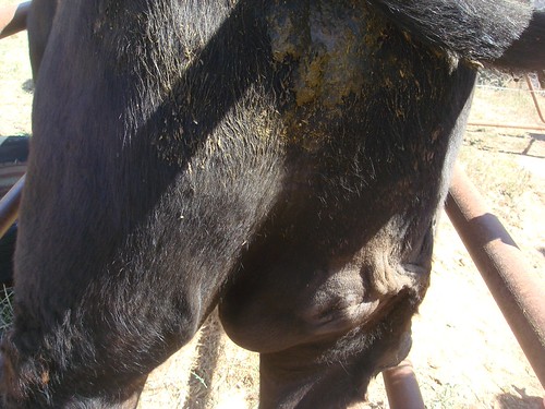
Cow with a swelling on its inner thigh. I probed it as I was concerned it was a migrating foreign body.
Ewe (female sheep) with an abdominal swelling. We anesthetized her and discovered this was a hernia.
Sophira’s eye lump.
Valvular lump in a very old dog. I desexed her and resected the lump and luckily she fully recovered.
4. If you leave a lump to grow too big on your pet, there is a huge chance it will burst. I strongly advise you to avoid this scenario.
This geriatric Labrador was in a really bad way when she arrived at the clinic. Her fatty lump had burst and was so infected.
Thankfully my colleague was able to resect the lump and close up her wound. She recovered really well.
5. Not every pet is going to be as lucky as the Labrador above. If you leave some lumps to grow too big, it is sometimes too late or impossible to remove them.
This geriatric terrier reached the point where he couldn’t defecate. His owners opted to put him down at this point. I was saddened to see he was left until he reached that point.
Sophira was 8 years old and very much loved. Her owners had brought her in earlier when the lump was much smaller. They were petrified about losing her under general anesthetic so they kept monitoring the lump. It reached this size and they decided it had to be removed. Unfortunately surgery didn’t go well as it was quite challenging to close the wound and she didn’t survive the anesthetic. Very sad outcome.
Sooty came to me 6 months ago and she had a small mammary tumor. It suddenly progressed and grew very quickly. We collected at biopsy and unfortunately we got inconclusive results and weren’t sure what we were dealing with at that stage.
We proceeded with a palliative surgery as she was quite uncomfortable from the weight of the lump. Unfortunately during the surgery, I discovered the entire abdominal wall was involved with the tumor and her abdominal organs were exposed.
She would have required major reconstruction surgery to close her wounds and there was a high risk of herniation. After speaking to her owners during the surgery, we had to put her down while she was still under general anesthetic. Absolutely heart wrenching outcome that may have been avoided.
6. Don’t assume the lump will outlive your dog. I often get owners thinking their dogs are too old to undergo surgery. They often don’t realize they are compromising their pet’s well being when they don’t address the lump.
Monty is a very old border collie that developed a very massive fatty lump. He was starting to really struggle to move around and so the owners decided they simply couldn’t put off this surgery any longer.
He felt like a brand new man after the lump was resected. It would have been far cheaper and a much short anesthetic if the lump was removed when it was smaller.
Jackson developed this lump over 1.5 months. His owners were quite concerned it was going to rupture and so in spite of cost constraints, they went ahead with the surgery.
He recovered brilliantly and we found out it was only a fatty lump. His owners are thrilled with the outcome.
Sorry for overwhelming you with so many gory pictures in this post but I hope it got the main message across.
I bet you are all feeling quite lumped out right now. Please fire away any questions you may have.
Related articles
- Oh no,I discovered a lump on my pet! Part 1 (rayyathevet.com)
Source: http://rayyathevet.com/2013/02/02/oh-no-i-discovered-a-lump-..
| Tweet |


Facebook Comments